Paper · Science Advances · 2020
Thousands of wires, one chip
Bundles of up to 8,640 fine wires were pressed onto camera, display and recording chips, then used to record neurons in retina and in awake, running mice.
Cancer Neurotechnology Lab
14 October 2026 · 10 min read
Silicon chips can hold hundreds of thousands, even millions, of tiny electronic pixels. The brain has a three-dimensional structure that flat chips cannot reach into. This study, published in Science Advances in 2020 by a collaboration between Stanford University, the Francis Crick Institute, ETH Zürich and others, connected the two with bundles of fine metal wires.[1]
The principle is the same as in the companion CHIME paper:[2] one end of the bundle presses against a chip, the other enters the tissue. This paper pushed the idea further in scale and in the range of chips and experiments.
A spacer that disappears
If you bundle wires too tightly, they enter tissue as one solid block rather than as many separate needles. The team solved this with a sacrificial layer.[1] Each insulated wire was coated with a plastic called parylene-C. The thickness of that coat sets the distance between neighbouring wires once they are packed together. The bundle was then filled with epoxy, and its top end polished flat for the chip.
At the brain end, oxygen plasma etched away the spacer and the epoxy, releasing the wires so each could penetrate individually. The free length of the wires could be set from half a millimetre to well over a centimetre, and the ends could be shaped, for example to reach the cortex and deeper structures at the same time.
The authors calculated how much tissue such a bundle displaces. For 15-micrometre wires spaced 100 micrometres apart, the arrangement used in mice, the wires occupy about 2% of the tissue volume they enter.
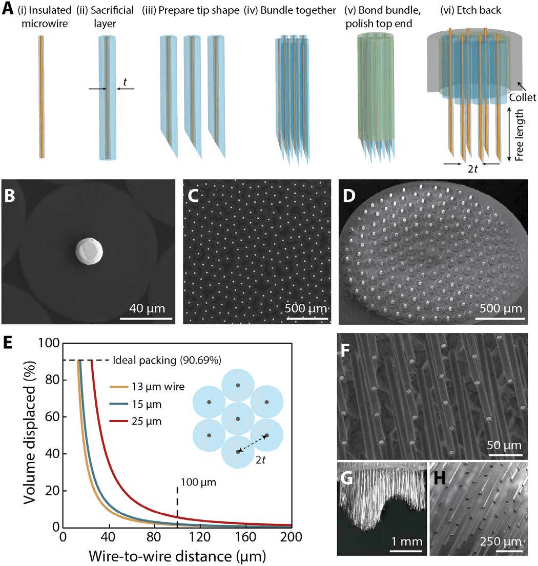
How the bundles are built. Wires are coated with a removable spacer, bundled, set in epoxy and polished, then released at the tissue end (A–D). The graph shows how wire spacing controls the fraction of tissue displaced: 2% for 15-micrometre wires 100 micrometres apart (E). Released wires can be cut to equal or varying lengths (F–H).
Reproduced from Obaid, Hanna, Wu, Kollo et al., Sci. Adv. 6, eaay2789 (2020), under CC BY 4.0.Pressing wires onto chips
At the chip end, the insulation was etched back to leave about 10 to 20 micrometres of bare metal on each wire.[1] A spring-loaded press then lowered the bundle onto the chip. As the bare tips touched the surface, they bent over and crimped against the pixels, forming electrical contacts even where the chip surface was not perfectly flat.
The connection was robust. The press alone kept connectivity unchanged over two weeks, and the interface survived being flicked with a finger.
The same method worked with three very different chips, chosen to show that it does not depend on any one design:
- the imaging chip from a Xenics Cheetah infrared camera, with 327,000 pixels;
- an OLED display chip from Olightek, with 1.44 million pixels; and
- a high-density recording chip (MEA) with about 25,600 pixels.
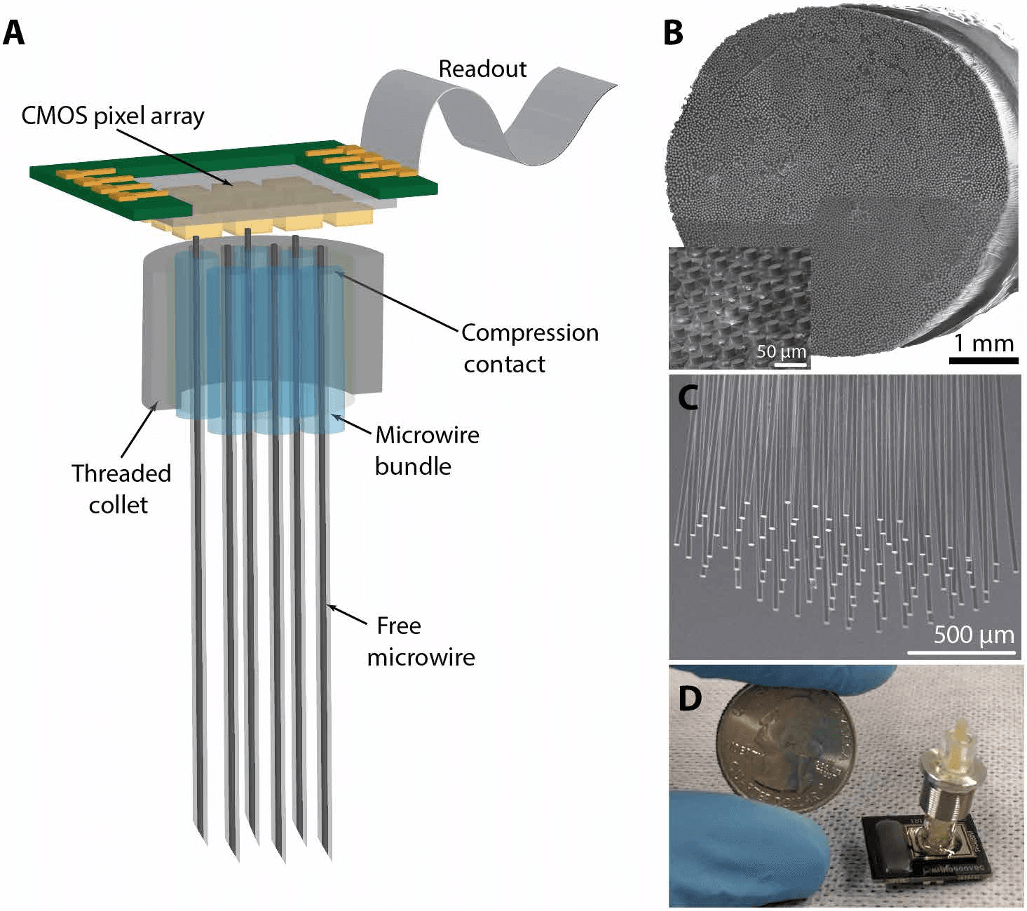
The design. A microwire bundle is pressed against a chip at one end (A, B) and splays into individually penetrating wires at the other (C). Panel D shows a bundle of 800 wires spaced 100 micrometres apart, in a device less than 0.6 cm wide.
Reproduced from Obaid, Hanna, Wu, Kollo et al., Sci. Adv. 6, eaay2789 (2020), under CC BY 4.0. Photo in panel D: Mina Hanna, Stanford University.How many wires connect?
Because the chips have many more pixels than the bundle has wires, the wires do not need to be lined up one-to-one with pixels. A wire may touch one pixel, or two, which simply gives two readings of the same wire.[1]
A 184-wire bundle connected 177 wires, a 96% yield. To show scale, the team pressed a bundle of 8,640 gold wires, about 7 millimetres across with wires 40 micrometres apart, onto the camera chip. More than 90% of the wires connected to an active pixel.
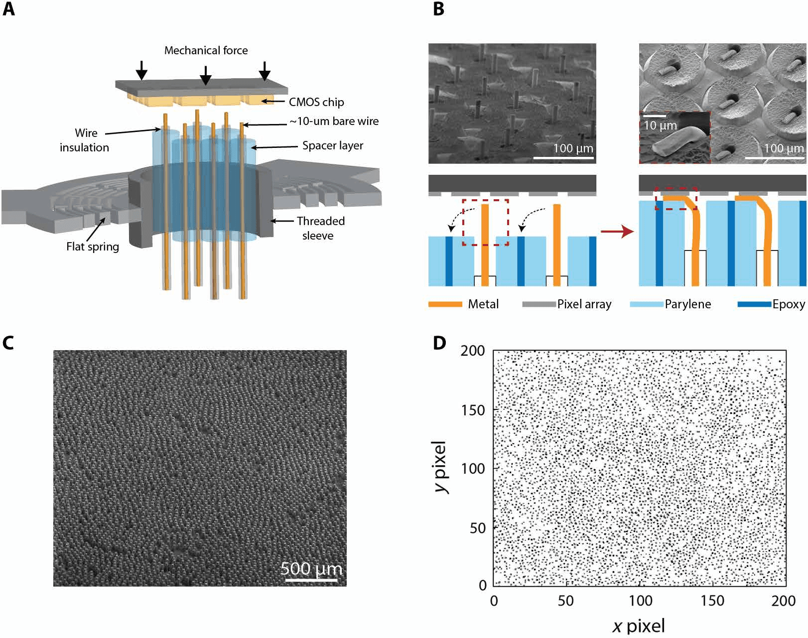
Contact at scale. A spring press lowers the bundle onto the chip (A), crimping the bare wire ends against the pixels (B). Panels C and D show part of the 8,640-wire bundle and its connectivity map on the camera chip, with more than 90% of wires connected.
Reproduced from Obaid, Hanna, Wu, Kollo et al., Sci. Adv. 6, eaay2789 (2020), under CC BY 4.0.Does the connection add noise?
Pressing thousands of metal wires onto a chip could, in principle, add electrical noise. The team measured noise on the recording chip before and after connecting a bundle of 251 platinum–iridium wires. It rose only slightly, from 5.0 ± 1.5 to about 6.0 ± 2.2 microvolts RMS, which the authors describe as well within the tolerable range for recordings in the brain.[1]
The chip connection also allowed a new trick. Using the OLED display chip, the team applied voltage to each wire individually to grow a coating of iridium oxide on its tip, a material that lowers impedance. Average impedance at 1 kHz for a bundle of 150 gold wires fell substantially after coating (Fig. 4C). The authors note this could let a single chip treat hundreds to thousands of wires in one step. For the animal recordings they chose platinum–iridium wires, because of their low impedance (200 ± 27 kilohms) and long-term biocompatibility.
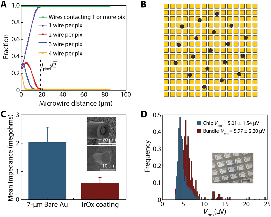
Connection, coating and noise. With enough spacing, every wire touches at least one pixel without alignment (A, B). Wire tips coated with iridium oxide through a display chip had lower impedance than bare gold (C). Adding a 251-wire bundle increased chip noise only slightly (D).
Reproduced from Obaid, Hanna, Wu, Kollo et al., Sci. Adv. 6, eaay2789 (2020), under CC BY 4.0.8,640
Wires in the largest bundle
over 90%
Wires connected in that bundle
5.0 → 6.0 µV
RMS noise, bare chip vs with 251-wire bundle
~2%
Tissue volume displaced (15 µm wires, 100 µm apart)
Recording from retina
The retina is a flat sheet of neurons, which makes it a good test for a device that samples a surface. The team placed a piece of isolated rat retina against a 138-wire bundle connected to the recording chip.[1]
They recorded 152 units in total, about 1.1 per wire, with signals more than 4.5 times the standard deviation of the noise. Firing increased at the onset and offset of a light pulse, as expected from the known ON and OFF types of retinal ganglion cells.
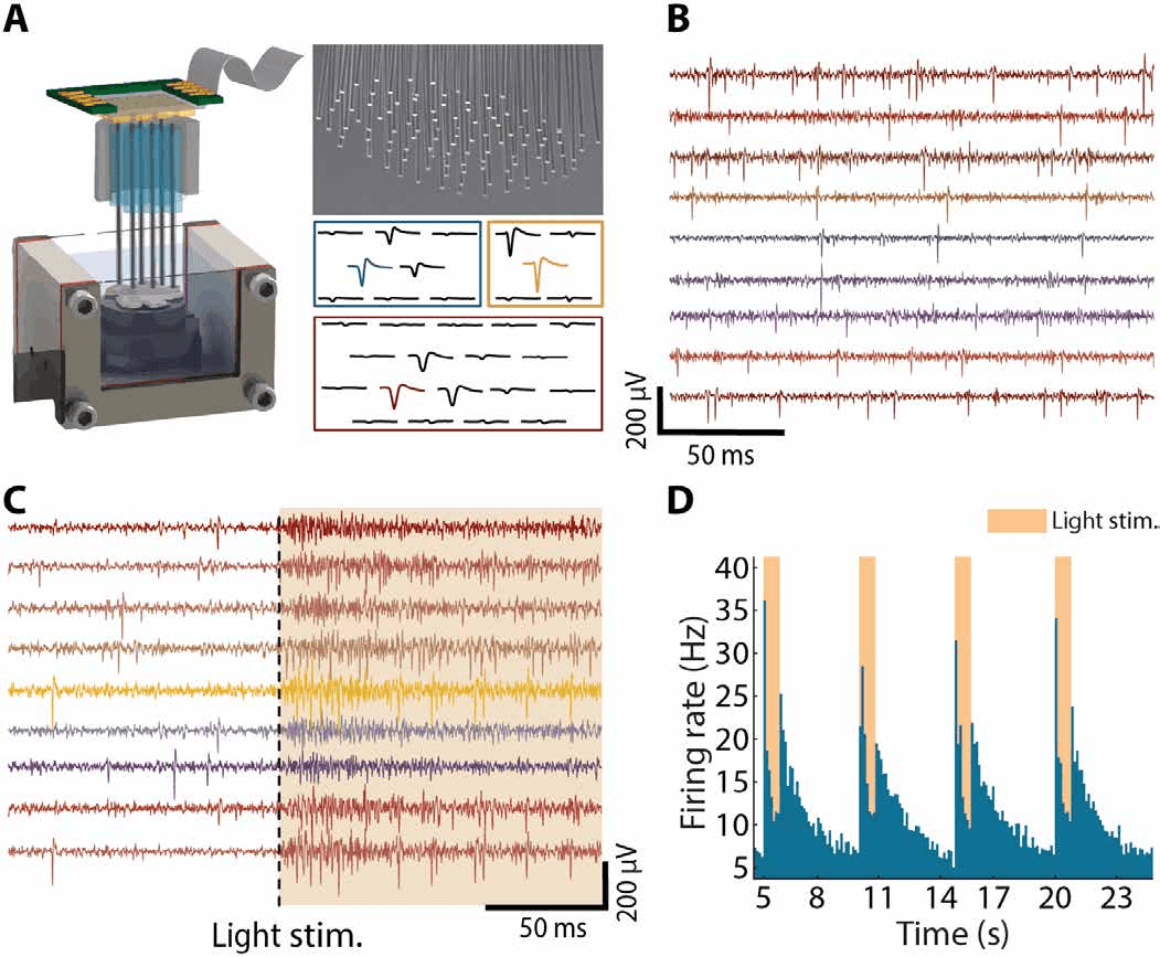
Retina on a wire bundle. Isolated retina rests on the bundle in a perfusion chamber (A). Wires recorded spontaneous activity (B) and responses to light (C), with firing rising at each light pulse (D).
Reproduced from Obaid, Hanna, Wu, Kollo et al., Sci. Adv. 6, eaay2789 (2020), under CC BY 4.0.Recording in awake, moving mice
The main test was in the living brain. Mice were head-fixed but free to run on a floating ball treadmill while bundles of 135 to 251 platinum–iridium wires, each 15 micrometres across and about 100 micrometres apart, recorded from the motor and somatosensory cortex or from the deeper striatum.[1]
Recordings were made within two hours of implantation. In each typical five-minute session, the team identified 92 to 221 putative neurons, about 0.56 single units per wire on average.
The recordings tracked behaviour. In the motor cortex, average firing rose from 8.4 spikes per second at rest to 20.1 while running. In the striatum it rose from 13.1 to 18.1. Gamma-band brain rhythms (30 to 80 Hz) in the striatum were also stronger during running than at rest, consistent with earlier reports.
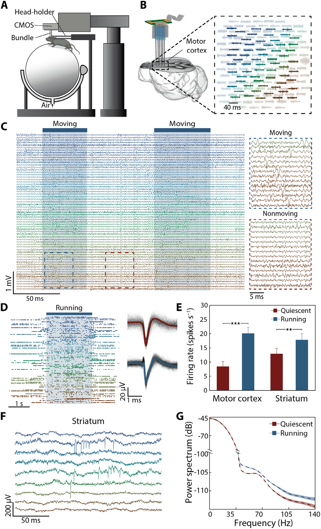
Neurons and behaviour. A head-fixed mouse runs on a treadmill while a bundle records across a broad area of cortex (A, B). Activity rises during running (C, D). Firing rates were higher during running in both motor cortex and striatum (E), and striatal gamma rhythms were stronger (F, G).
Reproduced from Obaid, Hanna, Wu, Kollo et al., Sci. Adv. 6, eaay2789 (2020), under CC BY 4.0.152
Units recorded in rat retina (138 wires)
92–221
Putative neurons per 5-min session in mice
8.4 → 20.1
Motor cortex spikes/s, rest vs running
13.1 → 18.1
Striatum spikes/s, rest vs running
What this does and does not show
- What was demonstrated. Bundles of insulated microwires can be connected at high yield to several kinds of commercial chip, without adding substantial noise, and used to record many single neurons in retina and in awake, moving mice.[1]
- What was not tested. Recordings were acute, made within hours of implantation, with the animal head-fixed. The authors describe miniaturising the device so animals can move freely as work underway at the time, and suggest that implantation under the skull may be possible given the chip's low power use (under 70 milliwatts). Neither was demonstrated here.
- Scope. This is a recording platform tested in animals. No experiments were performed in people.
Why it matters
The authors describe the approach as a third option alongside planar silicon probes and micromachined silicon arrays: it keeps the low tissue damage of small microwires while letting neuroscience use the latest silicon chips as they appear. Improvements in chip design, such as higher channel counts, stimulation or electrochemical sensing, could in principle be adopted by swapping the chip rather than redesigning the electrode.
For brain–computer interfaces, the paper shows a concrete path to channel counts that scale with the chip industry rather than with hand assembly.
Who did the work
Abdulmalik Obaid, Mina-Elraheb Hanna, Yu-Wei Wu and Mihaly Kollo contributed equally, with Romeo Racz, Matthew R. Angle, Jan Müller, Nora Brackbill, William Wray, Felix Franke, E. J. Chichilnisky, Andreas Hierlemann and Jun B. Ding. Andreas T. Schaefer and Nicholas A. Melosh share senior authorship.[1]
According to the paper's author contributions, the concept and design were initiated by Obaid, Hanna, Kollo, Racz, Angle, Schaefer and Melosh, and Racz was among the authors who performed experiments, acquired data and contributed to analysis and interpretation.
Competing interests, as declared in the paper. Three authors co-founded and hold shares in Paradromics Inc., a company developing scalable electrophysiology, and one author is a co-founder of MaxWell Biosystems AG. Patents related to the work were filed by some of the authors.
Animal procedures were approved by Stanford University's Administrative Panel on Laboratory Animal Care, as stated in the paper.
Sources
- [1]Obaid A, Hanna M-E, Wu Y-W, Kollo M, Rácz R, et al. Massively parallel microwire arrays integrated with CMOS chips for neural recording. Science Advances 6(12):eaay2789 (2020). Open access. doi.org/10.1126/sciadv.aay2789
- [2]Kollo M, Rácz RR, Hanna M-E, et al. CHIME: CMOS-hosted in vivo microelectrodes for massively scalable neuronal recordings. Frontiers in Neuroscience 14:834 (2020). Open access. doi.org/10.3389/fnins.2020.00834
How to cite the original work: Obaid A, Hanna M-E, Wu Y-W, Kollo M, Racz R, Angle MR, Müller J, Brackbill N, Wray W, Franke F, Chichilnisky EJ, Hierlemann A, Ding JB, Schaefer AT, Melosh NA. Massively parallel microwire arrays integrated with CMOS chips for neural recording. Science Advances. 2020;6(12):eaay2789. doi:10.1126/sciadv.aay2789
Figures are reproduced from the original open-access article under the CC BY 4.0 licence. Captions have been rewritten for a general audience; see the paper for the authors’ full captions.
This summary is written for public understanding and is not medical advice.