Paper · Journal of Neural Engineering · 2022
Listening to neurons without crowding them out
How glass-coated metal fibres finer than a human hair can record from, and stimulate, single brain cells while leaving the tissue around them largely undisturbed.
Cancer Neurotechnology Lab
14 October 2026 · 12 min read
Every recording of brain activity begins with a compromise. To hear what a neuron is saying, you have to put a sensor close to it. But the brain is soft, densely wired and threaded with tiny blood vessels, and anything you push into it takes up space that used to belong to living tissue.
A study published in the Journal of Neural Engineering in 2022 set out to make that compromise less costly.[1] The team, working mainly at the Francis Crick Institute in London with collaborators in Germany and elsewhere, built electrodes from continuously drawn glass-and-metal fibres and gave their tips a nanostructured coating. They called them jULIEs. The paper reports that these fibres recorded the electrical activity of single neurons, stimulated them, and did so with little detectable damage to the tissue around them, in experiments in mice.
Why size matters
The most common way to study how neurons work together is to record their electrical signals from outside the cells, using fine metal contacts called extracellular electrodes. These signals are fast. Neurons coordinate on the scale of milliseconds or less, and catching that timing is what makes electrodes such powerful tools for neuroscience and for brain–computer interfaces (BCIs).
The difficulty is that probes damage what they measure. The paper reviews earlier work showing that both immediate tissue damage and long-term deterioration track closely with the diameter of the probe, across many probe designs.[1] The obvious fix is to make probes thinner. But a thinner probe usually means either fewer recording channels or smaller metal contacts, and a smaller contact couples less well to the faint electrical signals around it. The signal shrinks and the noise grows.
jULIEs were designed to break that trade-off: keep the probe very thin, and fix the electrical coupling with chemistry at the tip.
Making a fibre finer than a hair
The fibres are made by a method first patented in 1931 and adapted here: a metal rod is placed inside a glass tube, the assembly is heated by induction to between 850 and 1,000 °C, and a thread of molten glass with a metal core is pulled off and wound onto a drum at about two kilometres per minute.[1] By adjusting the drawing conditions, the team could tune the metal core to between roughly 1 and 10 micrometres and the glass coat to up to 15 micrometres thick. Single fibres could be made in continuous lengths of more than 10 metres.
Microscope measurements showed that the glass surface is extraordinarily smooth, flat at the scale of nanometres. That matters because a smooth shank slides through tissue rather than catching on it.
The fibres were then bundled, set in a dissolvable resin, polished in bulk to a sharp 30-degree point, and freed again. Finally, they were bonded to small circuit boards to make probes with 16, 32 or 128 channels. The authors report that building and characterising a standard 16-channel probe took about 150 minutes, comparable to making a tetrode, a classic hand-built electrode.
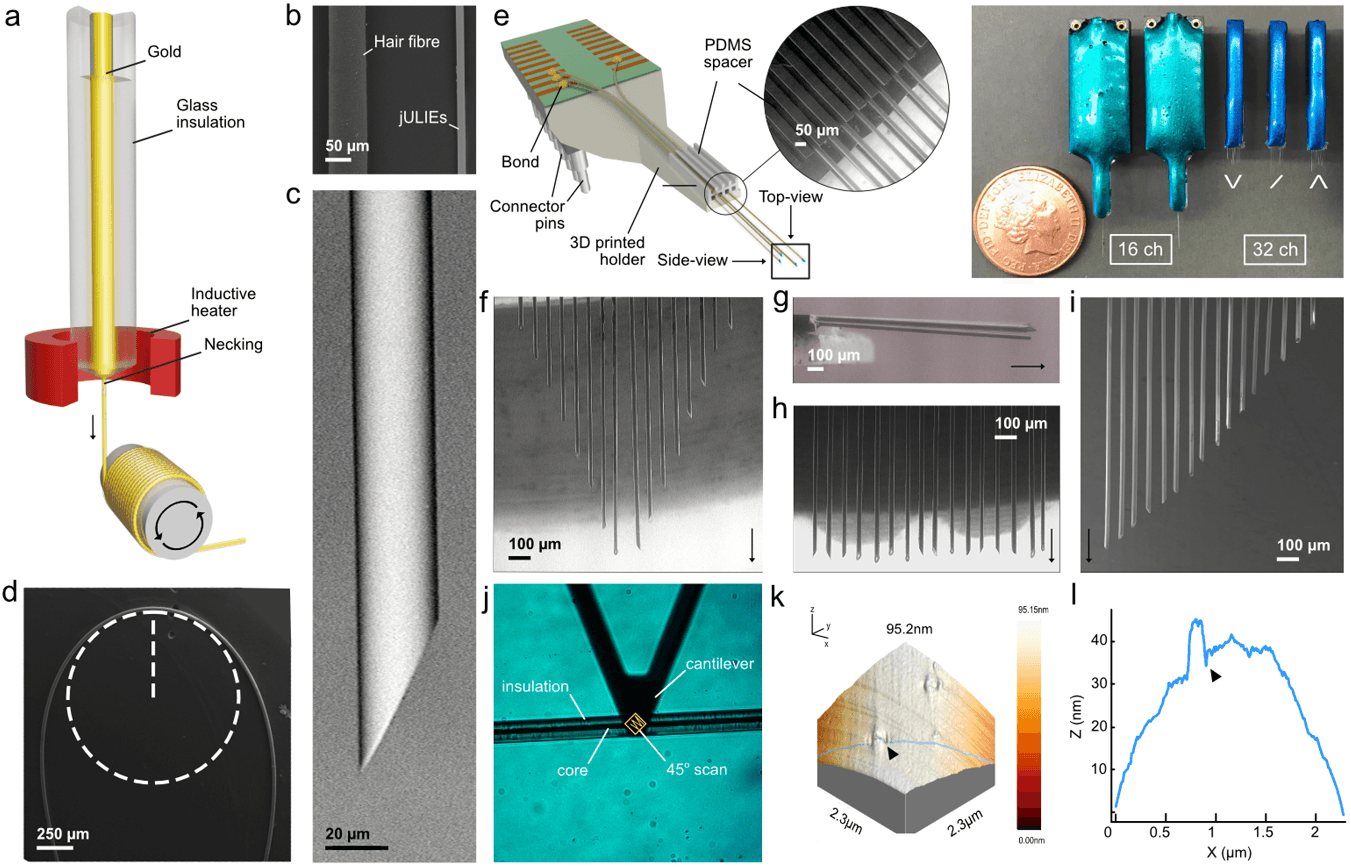
From molten glass to multi-channel probe. Fibres are drawn from a heated glass-and-metal preform (a), are far thinner than a human hair (b), and are sharpened to a point (c). They stay flexible (d) and can be assembled into probes with custom layouts (e–i). Atomic force microscopy shows a glass surface flat to the nanometre scale (j–l).
Reproduced from Rácz, Kollo et al., J. Neural Eng. 19 016041 (2022), under CC BY 4.0.Teaching a tiny tip to listen
Sharpening a fibre exposes only a small disc of metal at its tip, a few tens of square micrometres at most. A contact that small has a very high impedance at the frequencies used to record neurons, which weakens the signal and adds noise.[1]
The team's solution was to grow a rough, sponge-like structure on the tip, so that it has far more surface area without becoming much wider. They did this in two electrochemical steps. First, they deposited layered gold nanostructures onto the bare metal. Then they coated those with a thin film, about 20 nanometres thick, of iridium oxide, a material long used on recording and stimulating electrodes.
The gold layer has a second benefit. It seals the core metal away from the tissue, so the core can, in principle, be made from metals chosen for how well they draw into fine fibres rather than for how they behave in the body.
The result was a large drop in impedance. Measured at 1 kHz, a frequency relevant to neural spikes, the combined coating lowered impedance by up to two orders of magnitude, to around 100 kilohms, from a bare-tip starting point in the tens of megohms.[1] The coatings also held up: they survived insertion into deep brain structures, and after a million stimulation pulses the impedance had changed from about 200 to about 800 kilohms, which the authors note is still well within the useful range for recording.
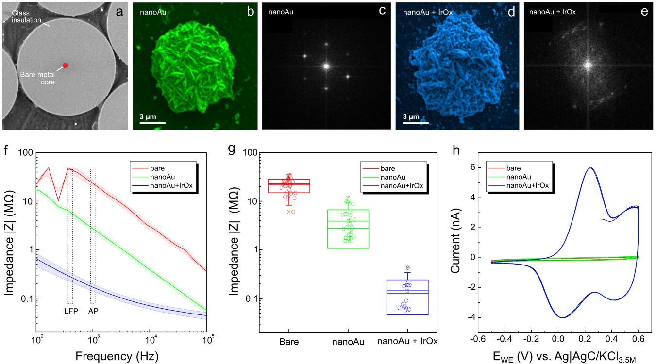
A rougher tip, a clearer signal. A bare polished tip (a) is coated with gold nanostructures (b, c) and then iridium oxide (d, e). Each step lowers the impedance across the frequencies used to record brain activity (f, g), and the fully coated tip can store and pass much more charge (h), which is what makes stimulation possible.
Reproduced from Rácz, Kollo et al., J. Neural Eng. 19 016041 (2022), under CC BY 4.0.Does the brain notice?
The most striking part of the study is what happened to the tissue.
Using two-photon microscopy in the olfactory bulb of anaesthetised mice, the team watched a fluorescently labelled fibre being pushed into the brain while blood vessels were also labelled. The paper describes capillaries folding around the fibre and sliding along its surface, without being dragged along or visibly torn.[1]
They then tested the blood–brain barrier directly. A blue dye that binds to a blood protein and normally cannot cross the barrier was injected into the bloodstream before a probe was inserted. Around a standard silicon probe, the dye leaked into the tissue, and cell nuclei were displaced and thinned out around the probe's rectangular shank. Around a three-fibre jULIE probe, the authors detected no leakage, and cell density looked undisturbed.
Function told the same story. The authors note that with silicon probes, neural activity is typically suppressed for up to about 45 minutes after a fast insertion, citing earlier work.[2] With jULIEs, single neurons could be recorded immediately, from the surface layers down. Some spikes exceeded 1 millivolt, a size which, the authors note, suggests the tip was very close to an intact neuron. When a fibre was pushed past a neuron and pulled back several times, the same activity reappeared at the same spot.
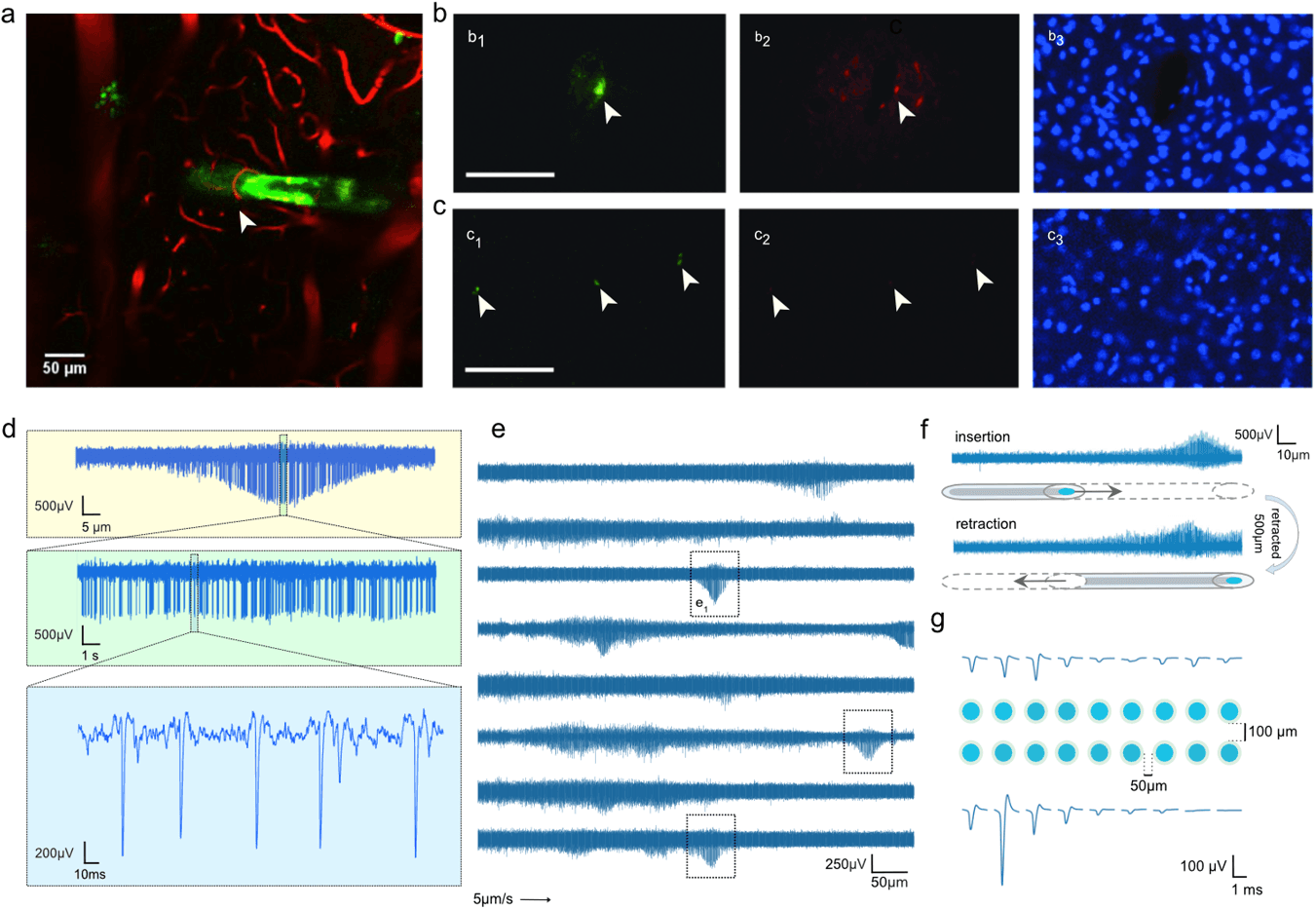
Sliding past, not pushing through. Blood vessels fold around a jULIE during insertion (a). A silicon probe causes dye leakage and displaces cells (b), while jULIE tracks show neither (c). Recordings made during slow insertion show individual neurons' spikes rising and falling as the tip passes them (d, e), and the same activity returns after repeated insertion and retraction (f). Neighbouring fibres pick up the same neuron (g).
Reproduced from Rácz, Kollo et al., J. Neural Eng. 19 016041 (2022), under CC BY 4.0.How close is close?
To estimate how near the tip got to the neurons it recorded, the team simulated the electrical field around a detailed computer model of a mitral cell, a principal neuron of the olfactory bulb. The model showed that the way a spike's size rises and falls as an electrode moves past a cell reveals how far away the cell is. Applying this to real recordings, they estimated that stable recordings were made as close as about 10 micrometres from the part of the neuron where spikes begin.[1]
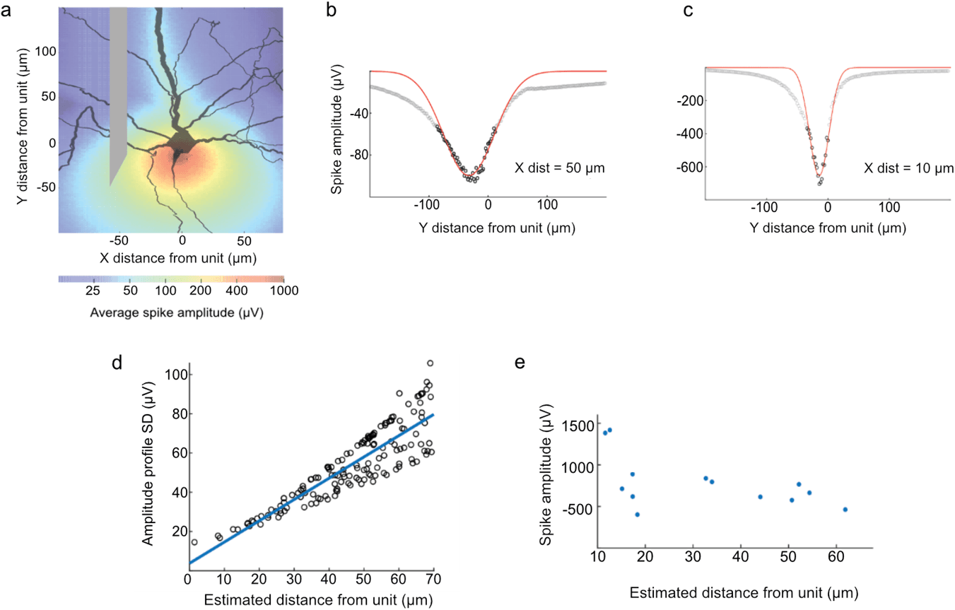
Reading distance from the signal. A biophysical model of a neuron (a) shows that the spread of spike sizes along the electrode's path depends on how far the path is from the cell (b–d). Applied to real data (e), this suggests recordings were made within about 10 micrometres of the neuron.
Reproduced from Rácz, Kollo et al., J. Neural Eng. 19 016041 (2022), under CC BY 4.0.Talking back: stimulation
Recording is only half of a two-way interface. The iridium oxide coating can store and release charge, which is what an electrode needs in order to stimulate cells.
To test this, the team used mice whose olfactory bulb neurons glow when they are active, and passed pulses of current through a single jULIE fibre while imaging the surrounding cells. Cells responded more strongly as the current increased, and most cells near the tip responded at 50 microamps.[1] The electrical properties of the tip stayed stable through 200,000 pulses, and after one million pulses the impedance had risen only three- to four-fold, so the same probe could still be used for recording.
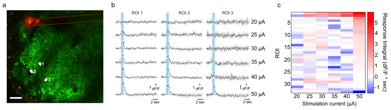
Driving neurons with a single fibre. Nearby neurons, made to glow when active, respond to current pulses from one jULIE (a, b), and the average response grows with stimulation strength (c).
Reproduced from Rácz, Kollo et al., J. Neural Eng. 19 016041 (2022), under CC BY 4.0.Shaping the probe to the brain
Silicon probes are made with chip-fabrication methods, so their recording sites usually sit along a flat shank, and changing the layout is a major engineering effort. Because each jULIE is its own fibre, the team could arrange them in custom two- and three-dimensional patterns, with fibres as close as 50 micrometres apart, to match the anatomy being studied.[1]
They showed this in two sensory areas.
Hearing. In the inferior colliculus, a midbrain hub for sound, different depths respond to different pitches. The team built a stacked, staircase-shaped probe to follow that layout. Each recording site picked up neurons tuned to their own preferred tone, and the probe recorded clear, tuned responses as little as about 40 micrometres below the surface, a layer the authors note is generally difficult to record from.
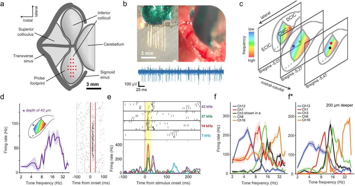
Following the brain's map of sound. A custom 16-fibre probe (a–c) recorded neurons with sharp pitch preferences, including close to the surface (d). Different sites preferred different tones (e, f), and the tuning held when the probe was moved deeper (f*).
Reproduced from Rácz, Kollo et al., J. Neural Eng. 19 016041 (2022), under CC BY 4.0.Vision. In the visual cortex, the team placed 16 fibres in a single row, 50 micrometres apart, at the depth of layer 5. They compared these recordings with separate recordings from a 16-channel silicon probe under similar conditions. The jULIEs had lower background noise, about 3.8 microvolts compared with about 12.9 for the silicon probe in this experiment, while the size of the spikes they recorded and the neurons' preference for the direction of moving stripes were statistically similar between the two.[1]
3.77 µV
RMS noise, jULIEs (n = 15 wires)
12.88 µV
RMS noise, silicon probe (n = 15 sites)
p = 0.28
Spike amplitude, jULIEs vs silicon
p = 0.88
Orientation selectivity, jULIEs vs silicon
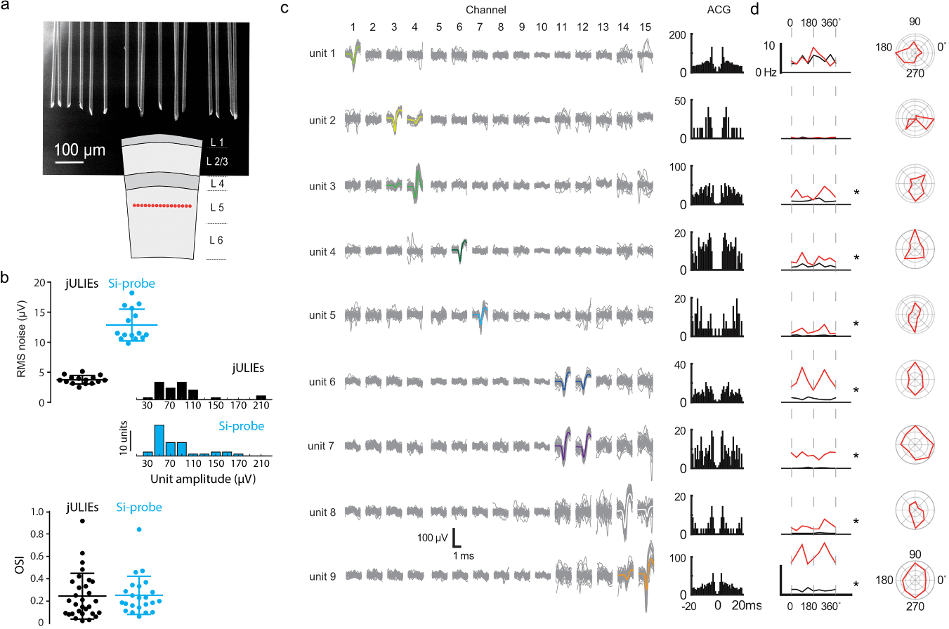
A row of fibres in one cortical layer. Sixteen jULIEs placed in layer 5 of the visual cortex (a) showed lower noise than a silicon probe, with similar spike sizes and tuning (b). Individual neurons were separated across neighbouring fibres (c) and showed distinct responses to moving visual patterns (d).
Reproduced from Rácz, Kollo et al., J. Neural Eng. 19 016041 (2022), under CC BY 4.0.Going deep. Finally, a 16-fibre probe was lowered step by step through the visual and somatosensory cortex, the hippocampus and the thalamus, down to the hypothalamus. Stable neural activity was recorded at every depth, to more than 6 millimetres below the cortical surface.
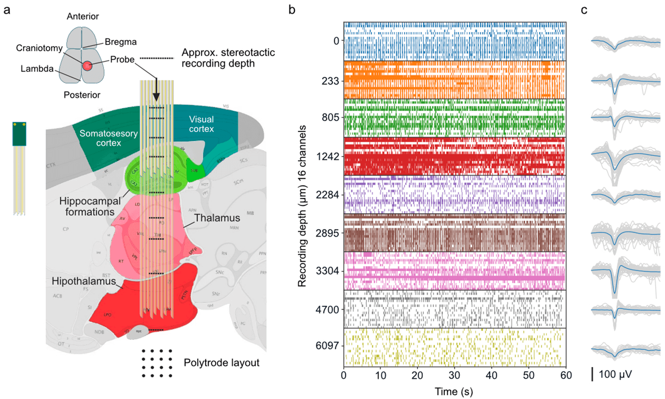
Top to bottom. A 16-fibre probe recorded neurons from the cortex down through the hippocampus and thalamus to the hypothalamus, more than 6 mm deep.
Reproduced from Rácz, Kollo et al., J. Neural Eng. 19 016041 (2022), under CC BY 4.0.What this does and does not show
It is worth being precise about the scope of this work.
- What was demonstrated. In acute experiments in anaesthetised mice, jULIEs recorded single neurons with low noise across many brain regions and depths, stimulated neurons, and caused no detectable blood–brain barrier damage in the tests performed.[1]
- What was not tested. The study did not test long-term implantation, which the authors identify as a key goal for future work. Electrical recording and imaging were not performed at the same time. No experiments were carried out in people.
- How it compares. The authors describe jULIEs and modern silicon probes as complementary. They give a sense of scale: a 20-micrometre microwire has a cross-section of about 314 square micrometres, compared with about 1,680 for a Neuropixels 2.0 silicon probe shank.
Why it matters
For neuroscience, the appeal is straightforward: a recording tool that changes the tissue less gives a more faithful picture of what that tissue is doing. The flexible layouts mean a probe can be shaped to the anatomy of a question, rather than the question being shaped to the probe.
For the wider field of neural interfacing and brain–computer interfaces, the study addresses the tension between miniaturisation and signal quality that runs through the whole field. Because each fibre has a metal core, the authors note that jULIEs can be connected directly to electronics, and at very large scale to flat silicon chips. That route was developed in related collaborative work on connecting microwire bundles to CMOS chips.[3][4] A patent naming some of the same inventors describes probes built from bundles of fine wire electrodes with impedance-reducing nanostructured tips.[5]
For our lab, this work is part of the technical foundation. The question we now ask is what living tissue can tell us at the boundary between a brain tumour and the brain around it. Measuring that boundary well depends on the principle this paper tested: get close to the cells, and change them as little as possible.
Who did the work
The paper's joint first authors are Romeo R. Rácz and Mihaly Kollo, with Gabriella Rácz, Ciprian Bulz, Tobias Ackels, Tom Warner, William Wray, Nikolai Kiskin, Chi Chen, Zhiwen Ye, Livia de Hoz and Ede Rancz. Andreas T. Schaefer is the senior author. The research was carried out mainly at the Francis Crick Institute, with collaborators at University College London, the Max Planck Institutes for Medical Research and for Experimental Medicine, Heidelberg University, Charité – Universitätsmedizin Berlin, the University of Southampton and others.[1]
The paper acknowledges support from the Francis Crick Institute's translation team through its Idea 2 Innovation grant scheme, and from funders including the Crick's core funders (Cancer Research UK, the UK Medical Research Council and the Wellcome Trust), the Human Frontier Science Program and the US NIH BRAIN Initiative.
All experiments were performed under UK Home Office regulations or German animal-welfare law, with approval from local welfare committees, as stated in the paper.
Sources
- [1]Rácz RR, Kollo M, et al. jULIEs: nanostructured polytrodes for low traumatic extracellular recordings and stimulation in the mammalian brain. Journal of Neural Engineering 19(1):016041 (2022). Open access. doi.org/10.1088/1741-2552/ac514fPubMed 35108701 · PMC7618266
- [2]Fiáth R, Márton AL, Mátyás F, Pinke D, Márton G, Tóth K, Ulbert I. Slow insertion of silicon probes improves the quality of acute neuronal recordings. Scientific Reports 9:111 (2019). doi.org/10.1038/s41598-018-36816-z
- [3]Kollo M, Rácz RR, Hanna M-E, et al. CHIME: CMOS-hosted in vivo microelectrodes for massively scalable neuronal recordings. Frontiers in Neuroscience 14:834 (2020). doi.org/10.3389/fnins.2020.00834
- [4]Obaid A, Hanna M-E, Wu Y-W, Kollo M, Rácz R, et al. Massively parallel microwire arrays integrated with CMOS chips for neural recording. Science Advances 6(12):eaay2789 (2020). doi.org/10.1126/sciadv.aay2789
- [5]Rácz R-R, Kollo M, Schaefer A. Electrochemical probe. US Patent 10,768,139 B2, granted 8 September 2020; assignee The Francis Crick Institute Ltd. patents.google.com/patent/US10768139B2/en
How to cite the original work: Rácz RR, Kollo M, Rácz G, Bulz C, Ackels T, Warner T, Wray W, Kiskin N, Chen C, Ye Z, de Hoz L, Rancz E, Schaefer AT. jULIEs: nanostructured polytrodes for low traumatic extracellular recordings and stimulation in the mammalian brain. Journal of Neural Engineering. 2022;19(1):016041. doi:10.1088/1741-2552/ac514f
Figures are reproduced from the original open-access article under the CC BY 4.0 licence. Captions have been rewritten for a general audience; see the paper for the authors’ full captions.
This summary is written for public understanding and is not medical advice.